New technic to treat eye disorder
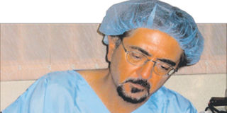
Dr Mukesh Joshi attending to a patient with a corneal problem. PHOTO/ISAIAH ESIPISU
After travelling for almost 5,000 kilometers to India, Kenyan Nihir Patel had to turn back because the treatment he was seeking was not available there then though he could have got it locally.
Patel visited India four years ago with an eye condition diagnosed as keratoconus – a degenerative disorder which can lead to blindness.
The condition usually affects teenagers. Here the cornea loses the normal shape of a watch glass and becomes very thin and steep resembling a cone and if not treated can even perforate making it totally white. Sometimes its thought to be hereditary or caused by particular allergies.


The latest treatment, Patel was advised by his doctors in India, is called cross linking but not available in India then and was referred to the Laser Eye Centre where the procedure was available.
300 eyes
In a study presented by Dr Mukesh Joshi, the medical director of Laser Eye Centre in Nairobi, at the 5th International Congress of Corneal Cross Linking last month in Germany about three hundred ‘Kenyan’ eyes have so far undergone the procedure.
“These are many young people with the most productive years ahead of them,” says Dr Joshi in an interview.
In cross-linking, Dr Joshi explains, the middle part of the cornea which is known as the stroma is strengthened by putting Riboflavin eye drops.
“This is followed by exposure to ultra violet light in such a way that is does not damage the rest of the eye, and is concentrated on the cornea.”
According to Dr Joshi it is difficult to diagnose keratoconus with the usual tools.
”The most dependable tool to diagnose the condition is called topolyzer.” In a topography examination concentric rings are projected on the cornea and images are taken by what is known as a CCD camera which are digitalised and converted on a flat screen that is colour coded.
Special
As thinning of the cornea is another sign of keratoconus, it is important to measure thickness of the cornea in the central and surrounding regions. This is done with a special ultrasound device known as a pachymeter or tomography
In the past, only optical treatment was available for keratoconus– where glasses or contact lenses were prescribed to the patient. This proves to be a difficult task as keratoconus patients do not have a very stable prescription.
Complex
The latest innovation in the treatment of keratoconus is what is called collagen cross – linking which was first initiated in Germany and Switzerland.
In cross-linking, Dr Joshi explains, the middle part of the cornea which is known as the stroma is strengthened by putting Riboflavin eye drops. This is followed by exposure to ultra violet light.
Complex calculations need to be carried out prior to the treatment so that the inner cells of the cornea are not damaged. Cross – linking makes the corneal stroma several times stronger.
If keratoconus is at a very advanced stage, then corneal transplant is recommended. The latest corneal transplant technology is called deep lamellar keratoplasty.