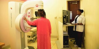
Vivian Mbaabu, a mammogram technologist, doing a mammogram at Aga Khan University Hospital.
It is 11am and I’m seated outside a breast imaging clinic at Aga Khan University Hospital. Two elderly women enter and exit within minutes—10 minutes each, to be precise. I had contemplated not coming for this mammogram, an X-ray examination of my breasts. Such things can be scary. I try to read the women’s faces to gauge whether the process is painful. They look calm, but I’m feeling queasier with each second.
For most women, myself included, the first thing we think of when doing a mammogram is cancer – and that thought elicits nothing but fear.
I had postponed this routine breast screening for four years, but this October, I committed to proving that I can regain control of my breasts. I knew that as a woman, I need to know what my inner breast looks like. I realised that most of us are concerned about our breasts for their sexual potency; we rarely give them the same attention we give our face, hair or skin.
As I approach the registration desk, the nurse hands me a questionnaire. I fill it and notice that perhaps my “yes” ticks are too many. I ticked yes to having breast pain and yes to lumps. I am among the women whose breasts are a source of torturous pain a few days pre-menses, during ovulation, peri-menopause, or any other day when our progesterone hormones go berserk. Women who have constant confusion about our cyclical breast changes.
At 11.26am, I walk into the mammography room. I stand with my chest exposed. The technologist, Vivian Mbaabu, asks me to place my breast on the machine. I am a size 34C, but I struggle to fill the whole breast onto the machine. A section of my breast spills out. Do women with bigger boobs find it easier to place their breasts on the plate-like machine? I wonder, but sensing this might be a stupid question to ask in a mammogram clinic, I let my mind drift back to my spilling breast. Vivian gently scoops it back and packs it in nicely. She asks me to lean forward and not move. Then the machine starts whirring as it presses the breast flat.
Interestingly, it is not uncomfortable. Within seconds, Vivian is done. She switches to the right breast.
After 10 minutes, I was done. She tells me that the mammogram had shown I had a few cysts, and the breast radiologist would explain more. My mind races, wondering if something ominous was brewing underneath these breasts, my marker of femininity. I knew that I had on-and-off cysts and lumps—non-cancerous—but I am a worrywart. I also have had three lumps surgically removed on the left breast, just because I didn’t want them haunting me.
It is not uncommon for women who have a screening mammogram to be requested to do additional imaging, according to Dr Rose Ndumia, the section head of breast imaging and vice-chair of the radiology department at Aga Khan University Hospital. Neither is it uncommon for women to have breast pain and lumps. Not all these women will be found to have cancer.
“There are findings such as benign lumps, cysts, and lymph nodes that just need a second look through a breast ultrasound. Also, lumpy breasts tend to hide small masses that can be seen clearly using breast ultrasound,” she says.
“Also, when the image gets into the hands of the radiologist, she may feel they didn’t get a good enough look at a certain part of the breast or some of the breast tissue wasn’t flattened out to read the mammogram properly.”
Because I am in the category of women with dense breasts that can obscure small masses, I was scheduled for a breast ultrasound.
As I lay, breast exposed, hands behind my head, on the examining bed, the ultrasound technologist uses a small device to search for the walnut-sized lump seen in the mammogram. A stick-like device roams up, down, and around, gently pressing into the breast and under the armpit. The ultrasound technologist, Victoria Khamati, repeats on the right breast.
I am dry-mouth-anxious, face turned away from the ultrasound screen, saying a silent prayer. I was lucky to have Dr Ndumia in the ultrasound room, as the technologist did the tests. Perhaps sensing how tense I was, she immediately says, “So far nothing to worry about, just cysts and a lymph node.” I sigh in relief.

Vivian Mbaabu, a mammogram technologist, doing a mammogram at Aga Khan University Hospital.
Lifecycle of breasts
The problem with breasts is that more than any other organ, they are constantly changing. Age, day of the month, pregnancy, breastfeeding and peri-menopause give rise to a worrying amount of pain, secretions, lumps and bumps, all of which can be normal or abnormal.
Cysts are common in women in their 30s to 50s. They are fluid-filled sacs. They can be achy. They can be many or clustered in one or both breasts. They may disappear, or a doctor may draw off the fluid using a syringe.
Lumpy breasts or not, every woman must do an annual breast screening.
“But you cannot know if it is a cyst or a non-cancerous lump by just touching it, you must do a breast ultrasound,” Dr Ndumia says.
She argues that both lives and costs are saved by doing yearly screenings. For women over 40, there are benefits to having your mammogram done in one hospital.
She shows me mammograms of women who have been routinely coming for years at the Aga Khan breast imaging clinic, which has a digital mammography machine that provides an electronic image that is stored as a computer file. The files are in hundreds, if not thousands and here, there are some women who have religiously been doing their mammograms every year.
“When someone has had prior mammograms in the same hospital, we can look back to your mammogram reports in the computer file and say, ‘Oh, this cyst or lump has been there for two years, and it hasn’t changed. We don’t need a biopsy,” Dr Ndumia says.” But if you come to this clinic, and last year you were in another clinic, we don’t have your records, so we don’t have anything for comparison.”
“Additionally, our machine does something called tomosynthesis, which you won’t see in all clinics. It produces a more detailed image of your breast than a standard mammogram,” she says.

Dr Rose Ndumia who heads the breast imaging section at Aga Khan University Hospital.
Stage zero
To show how significant early detection is, Dr Ndumia cites the difference between a small incision or the loss of breasts; the difference between a pill every day or months of chemotherapy and radiotherapy which have long-term side effects; the difference between stage zero diagnosis, or a life-threatening stage four prognosis.
“Look at this woman’s report. All her previous ‘mammos’ were clean, then we realised a change. Can you see the calcifications [deposits like grains of salt in the breast]? We biopsied it and it turned out to be cancerous but in the very initial stages. She only got a small breast incision to remove the cancerous tissue, no chemotherapy, no radiotherapy. She didn’t lose her entire breast.
How does the surgeon know where to operate on the breast? There’s something I normally do with the surgeon. I put a magnetic tape around the area [inside the X-ray showing cancer.] Then the surgeon will go in with a magnetic reader. When he gets close to that magnetic marker, he knows where he’ll cut out the cancerous tissue. On the day of the surgery, the patient’s husband was sent to bring me the specimen to check that they’d removed the whole cancerous tissue. We checked and the patient’s cancer had been scooped out.
“That means if she hadn’t come, the cancer would have spread. Had she skipped her routine screening, maybe it would have become a lump. The lump would have been an invasive cancer, which has the likelihood of spreading. That’s why mammograms are very important,” says Dr Ndumia.
A mammogram costs Sh2,950 and, combined with an ultrasound, it costs Sh8,500. If the disease is caught early, a small incision called a lumpectomy would cost between Sh90,000 and Sh300,000 while a double mastectomy plus breast reconstruction can cost up to Sh1.8 million in private hospitals. Chemotherapy including insertion of a chemo port can amount to Sh7 million.
To know if it’s cancer or not, Dr Ndumia, the only breast radiologist now working in Kenya, says your test results are as good as your radiologist.
“You need the right people to do breast imaging, the right equipment, and the right radiologist to interpret. In radiology, we have subspecialties. I’ve trained in radiology and breast imaging. In Kenya, we used to be four. The first person who trained with us abroad came back, practised a little, and then they moved to an international organisation. She was like, Rose, I have left this to you. Two of my colleagues went and trained in the UK. They have finished and are now working abroad. We lose a lot of brain power to other countries,” says the assistant professor who is now training more breast radiologists in Kenya to bridge the gap.
Her concerns over the not-so-experienced radiologists reminded me of my 2020 breast ultrasound procedure. A young radiologist, perhaps in his late 20s, saw something in my breast and started sweating. I looked at him in disbelief. ‘What have you seen,’ I asked him, my heart racing. His voice shaking, he replied, “I’m not allowed to tell you what I’ve seen, your doctor will tell you.”
Angry, and in between sobs, I asked him to call his supervisor. The supervisor requested a core biopsy, the most painful breast test I have ever done because they pushed the needle deep inside the breast tissue, without anaesthesia. My breast was sore and discoloured for weeks. Days later, the radiologist called back and said he did not pick enough cells, so I repeated the painful, and expensive test that had cost me Sh28,000.
“That’s why we need more breast radiologists. A general radiologist may interpret mammograms or breast ultrasounds, but when faced with challenges or something in finer detail, they need someone they can consult,” Dr Ndumia says. “If you go to a hospital with a poor-quality mammography machine, it might miss something. If the technologist is not well-trained, they might miss the cancer too.”
Family member
Her other concern is that Kenyans assume if no family member has had breast cancer, then screening is unnecessary. The Gail Model or the Breast Cancer Risk Assessment Tool can help one estimate the risk of developing breast cancer, but even if the risk is low, Dr Ndumia says annual screening is important.
“Even if you don’t have a family member who’s had breast cancer before, do screening, because even those with a family history of breast cancer, it’s only 25 per cent who will develop the disease. It’s not like a sure bet that because someone in your family had it you’ll get it,” she says.
At about midday, my mammogram and ultrasound visit had come to an end. My breast is classified as “heterogeneously dense.” I have less fatty tissue and more fibrous tissue in my breast than other women who do not have dense breasts.
“There are four categories of breast density,” Dr Ndumia says. For those with lumps, she says, follow-up check-ups are important.
“When you have solid mass, it is oval, and it is not worrisome, we ask you to come for a follow-up in six months, then another six months. if it remains stable for two years, we label it as benign [non-cancerous],” Dr Ndumia says.
There is this fear that a mammogram is painful, but it is not. It is also not as hazardous. “A mammogram has a low dose X-ray so you won’t get a lot of radiation,” she says.
Why can’t one skip a mammogram and do an ultrasound? I ask.
“Because of calcification [small white spots on mammograms which can be seen as randomly scattered, or appear in groups or as specks in a line.] Look at this patient’s mammogram report [she shows me]. She had no lump at all. She came because she had a relative who had breast cancer. From the mammogram, the doctor saw her breast had calcifications. You cannot see calcifications on an ultrasound,” she says.
If cancerous...
Doctors use a standard system to write mammogram findings and results, numbered BI-RADS 0 through BI-RADS 6. If your mammogram shows suspicious lumps, cysts or lymph nodes or calcifications, the report will be written “BI-RADS 4” or “BI-RADS 5”.
“A woman in such a case will be sent for a biopsy [where a needle is inserted into the breast and picks tissue for testing.] If positive, one is sent for a CT scan to stage the disease and to show if the cancer has spread to other parts of the body such as the lungs. After that, we discuss the treatment. Some patients will start with surgery [removal of the whole breast or part of it] and then do chemotherapy. Other patients will start with chemotherapy to shrink the tumour, then do surgery,” she says.