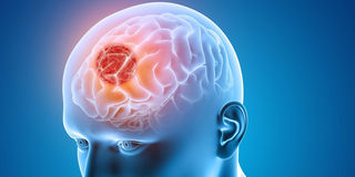
What you need to know about brain tumours
There is no one-size-fits-all treatment in brain tumour care and your plans should be discussed at length with your treating neurosurgeon.
What you need to know:
- 4.3 people in every 100,000 are diagnosed with brain tumours each year
- If the tumour is determined to be cancerous, radiation therapy may be required
- Treatment for metastatic brain tumours is very much guided by the specialists in the organ of origin of the tumour
Being diagnosed with a brain tumour can feel very stressful and isolating at the same time.
To many people, a brain tumour is this mystery disease that we would rather not discuss. Often, I meet people who equate this to cancer, which to most equals a death sentence.
In this article, I will try and unpack the basic facts about tumours. The subject is very wide and we certainly cannot cover it all comprehensively. We mark the World Brain Tumour Day on June 8.
What is a brain tumour?
A brain tumour is a collection of abnormal cells that no longer respond to the body’s mechanisms to kill off defective cells. We describe brain tumours as primary or secondary. Primary brain tumours originate from the brain cells (astrocytes or oligodendrocytes),organs associated with the brain such asthepituitary orpineal and the brain covering tissue (meninges). Secondary brain tumours are those that have cells that originate from a different organ with the most common sites being the breast, lung, prostate, large bowel (colon), kidney and skin. Secondary brain tumours are often referred to by the medical term metastasis.
Primary tumours are graded according to how much their cells have genetically changed from their original cell lines. This molecular genetic changes allow them to multiply much faster, invade nearby tissues and recruit blood vessels to aid their growth.
This changes sometimes allow them to camouflage themselves from the body’s systems to destroy normal cells and sometimes they aid the cells to invade other regions of the brain, spine or other body organs (metastasis).
The grading is 1-4 with 1 being the least changed and 4 being the tumour with the most molecular changes, making it more aggressive. Grade 1-2 are described as more benign while 3-4 are more aggressive or malignant. The grading of tumours is done on tissue and hence the discussion with your medical team to sample the tumour (medically referred to as abiopsy). The grading system informs your management, treatment options and future outlook (prognosis).
How common are brain tumours?
We do not know for sure the overall number of people who have brain tumours in Kenya because not everyone has access to a health professional who can make the appropriate diagnosis.
Secondly, we do not have a comprehensive central database that keeps track of all patients with brain tumours.
However, we can use statistics from other countries, to estimate how many patients we might expect to see. In the Global Burden of Diseases Study of 2016 published in the Lancet in 2019, they estimated that 4.3 people in every 100,000 are diagnosed with brain tumours each year.
With a population of 47.5 million at the last Kenyan census, that would give an approximate number of 2,200 people per year who get a new diagnosis of a brain tumour, or 42 people per week or 6 people per day. Brain tumours occur typically in childhood and then a second peak in later life.
How do they present?
Brain tumours present in several ways:
- General increased pressure in the skull resulting in a headache which increasingly does not improve with painkillers. The pressure can make someone nauseous or even vomit. Increased pressure can reduce someone’s conscious levels initially with agitation, confusion and eventually in coma.
- Disruption of brain pathways causing weakness, change in sensation, reduced ability to make decisions, loss of bladder control, speech difficulties, loss of hearing, sense of smell or vision. Other changes include incoordination, poor balance and repeated falls.
- Developmental regression is when someone loses the ability to perform functions they used to do. This description is mainly used in babies and children who may stop walking, stop talking or reduce their social interaction depending on the area of the brain that has been affected.
- Seizures which is essentially a short-circuit of the brain’s electrical conduction.
- Hormone imbalance is a marker of pituitary tumours. This can affect our metabolism, immunity, bone and muscle growth, sexual libido, fertility, breastmilk production and even our ability to wake up and have the energy to work all day.
How will I be investigated?
All doctors will want to hear about your symptoms (taking a history) as well as to examine you. Often, this can give us a very accurate picture of where in the brain you may have a problem but in itself cannot tell us exactly what has caused the problem.
As the brain is concealed in the skull, imaging is the next most useful test. A CT (computed tomography) uses X-rays to build a 2D or 3D image. A CT scan is often fast and most people can tolerate it. It is also much more available in the country and is therefore a very useful first test especially when the patient is not very stable and may need emergency treatment.
The next test is an MRI (Magnetic Resonance Imaging) which uses magnets to generate images of your brain. It is a much more detailed scan which helps predict with greater accuracy what type of tumour it could be. It is for this reason that if you are required to remain under surveillance with repeat imaging, you will often be asked to do an MRI. Although they are increasingly becoming available, not all scanners have the appropriate strength or sequences for treatment planning. Speak to your medical team before booking an MRI to ensure you have gone to the appropriate imaging centre capable of producing the required images.
Other imaging modalities such as PET (Positron Electron Tomography) imaging are useful in calculating tumour burden or spread especially in metastasis/ secondary tumours. Sometimes they are used to assess the metabolic activity in tumours after radiation therapy.
What are the treatment options?
There is no one-size-fits-all treatment in brain tumour care and your plans should be discussed at length with your treating neurosurgeon. Treatment depends on the imaging findings, location of the tumour, your age and your general fitness. Hormone imbalances and seizures respond very well to medical therapy which will be adjusted accordingly.
Small benign lesions that were picked up incidentally when being investigated for something else may initially be managed with surveillance with repeat imaging. Some of these lesions may not have any appreciable further growth and may therefore not need further treatment.
To establish the exact tissue type and to work out the molecular genetic changes, a sample of the brain tumour will need to be obtained. If the tumour is deep or in an area of sensitive brain mechanisms, a sample can be obtained with special navigation equipment (stereotactic biopsy) with a tube as thin as a strand of spaghetti, or a biro ink tube. If the tumour is accessible surgically you may have a surgery with the intention of taking out as much tumour as can be obtained without giving you new defects.
To aid this process, sometimes we use real time neuromonitoring tracking the brain and spinal cord electrical activity and sometimes we do the procedure with the patient awake, asking them to perform certain tasks that match the area of tumour being operated on.
Awake surgeries are quite a specialised skill requiring a specially trained team of staff led by the neuroanaesthetist (special doctor who puts patients undergoing neurosurgeries to sleep) as they need to balance your comfort, pain control and ability to respond to the surgeon.
Radiation and chemotherapy treatments
Once the tissue has been analysed and the tumour is determined to be cancerous, radiation therapy may be required. The radiation treatment plan is guided by an oncologist. They also guide any chemotherapy treatment indicated.
Sometimes chemotherapy and radiotherapy is indicated for treated tumours that recur or if there is remaining tumour tissue that was not surgically accessible. Most patients in this phase of treatment are seen in the outpatients making it easier for you to spend time with your family.
Treatment for metastatic brain tumours is very much guided by the specialists in the organ of origin of the tumour. For example, the input of specialists in breast cancer, is essential in managing patients with a tumour that originated from the breast cancer as they have the most comprehensive knowledge of the tumour’s genetic makeup and current treatment options.
For these reasons, most hospitals run a tumour-board which allows for collective treatment plans with different specialists advising. At the Aga Khan University Hospital, Nairobi, for example, we have tumour boards covering different types of cancers. This is particularly useful when a tumour type is rare, where new treatments may be available or where patients have had variable responses to various treatment strategies.
Can I be treated in Kenya?
The neurosurgical community in Kenya is growing with 37 in practice and counting. Many major cities in the country now have a neurosurgeon with more in training locally and abroad. Many of the treatments are available though not all of them are available in each centre.
Where you may benefit from a review in a different treatment centre locally or abroad, your neurosurgeon should be in a position to advise. Certainly, the techniques discussed above are available at the Aga Khan University Hospital, Nairobi.